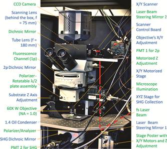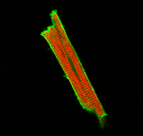
Two-photon and second harmonic generation confocal microscopy (SHG-TPEF) A hybrid SHG-TPEF polarization imaging system (Fig. 1) has established in our lab, with the excitation beam from an fs Ti:Sappire laser being bi-directionally raster scanned across the focal plane using a pair of orthogonal galvanometers. To implement high speed scanning, the turning regions of the triangle waves were smoothed by a custom-designed waveform. The SHG and TPEF signals from samples were recorded by two PMTs in the forward and backward direction respectively. Using the imaging system, we obtained 3D images of the sarcomere structure via SHG and DiO stained lipid membrane via TPEF in live cardiomyocytes isolated from neonatal and adult rats (Fig. 2). The results showed the potential applications of SHG and TPEF in the research of myofibrillogensis |
Fig. 1 Schematic of the SHG-TPEF hybrid microscope |
|
| Fig. 2 2D image of a freshly isolated adult cardiomyocytes: A) TPEF image of a live myocyte with DiO-stained membrane (for better color contrast, the DiO-stained cell membrane was displayed in green); B) simultaneous SHG image of the same cell, showing sarcomeric structure (red); and C) Combination of images A and B (Scale bar: 10 μm). |
|
|